
Objective: Aortic valve replacement in children is challenging. The lack of accommodation for somatic growth negatively impacts long-term outcomes. Data on the use of symmetrical oversized leaflets in neocuspidization are limited. We present a computer-based simulation of patient growth after a replacement with an oversized aortic valve leaflet.
Methods: A new aortic valve design was modeled in silico. Three computer-simulated symmetrical leaflets, each 18mm in nominal width, were constructed for 4 simulated aortic roots with different diameters (12, 14, 16, and 18mm. Pressure was applied to the aortic root side to simulate diastolic pressure and valve closure. The morphology of the closed valve was evaluated across different settings to examine morphologic changes during somatic growth. Three parameters of oversizing, previously described in our in vitro and in vivo studies, were assessed across the simulations: windmill shape deviation, coaptation length, and billow below the annular plane distance.
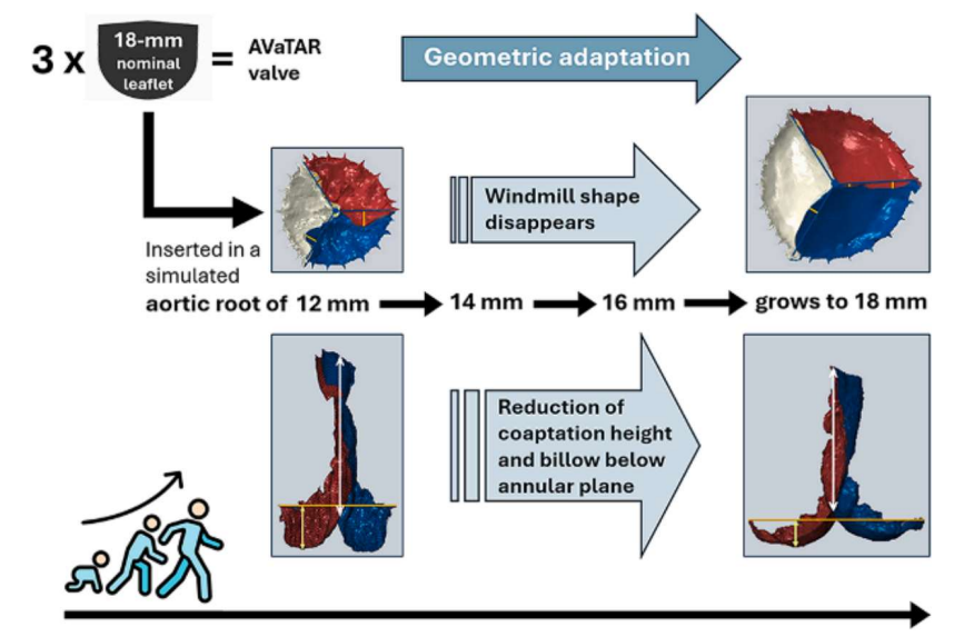
Results: A decrease in oversizing parameters was observed as the diameter of the aortic root increased. The average deviation from the windmill shape, the coaptation length, and the distance from the annular plane to the leaflet billow all decreased consistently with growth, indicating morphological adaptation of the valve geometry.These correlated changes suggest that the valve design adapts to anatomical growth by altering its functional configuration without compromising coaptation.
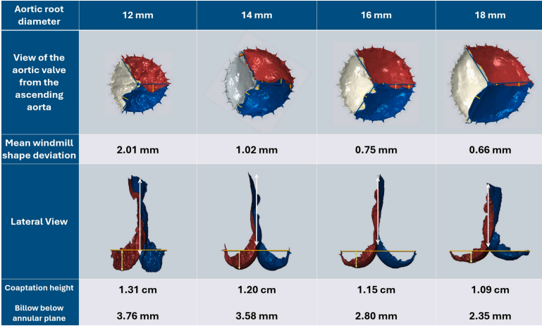
Figure: Evolution during growth of the 3 features that characterize oversizing of the aortic valve trileaflet autologous reconstruction valves.
Conclusions: As the aortic root enlarged from 12 to 18 mm, windmill shape deviation, coaptation length, and billow below the annular plane progressively decreased, indicating morphological adaptation of the valve geometry during growth.
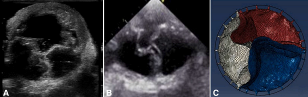
Figure: Morphologic similarities between in vitro (A) and in vivo (B) echocardiographic evaluations from our previous studies and the in silico simulations (C) performed in the present study.
Conclusions: The morphologic correspondence between the in vitro, in vivo, and in silico evaluations supports the validity of the computational modeling strategy.
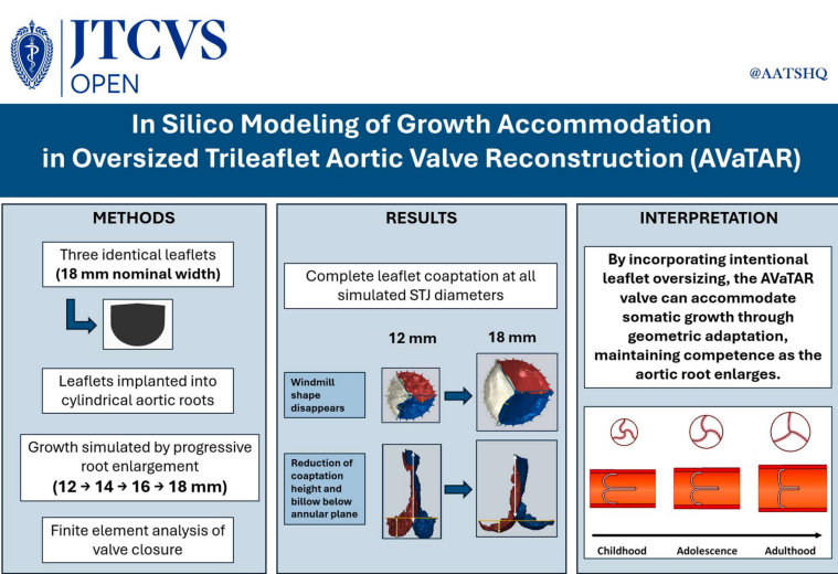
Figure: Geometric adaptation of the oversized AVaTAR aortic valve during somatic growth.
Conclusions: The oversized AVaTAR valve maintained complete leaflet coaptation across all simulated growth stages, supporting somatic growth accommodation through geometric adaptation as the aortic root enlarges.


.png)